


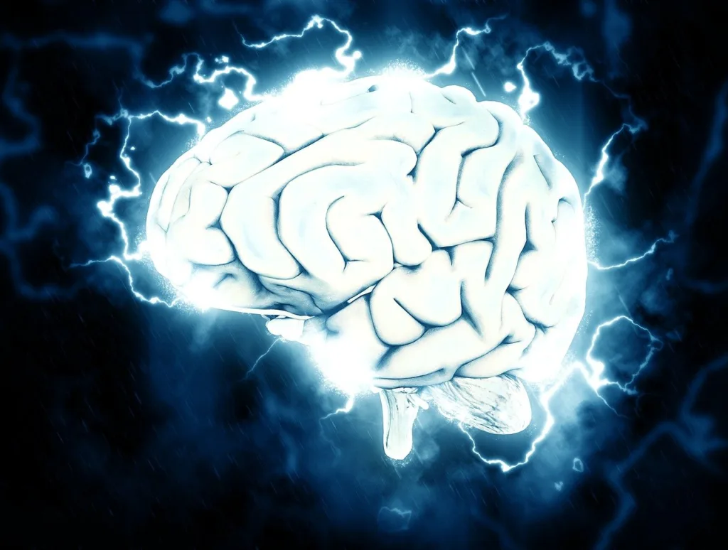
The oxygen pathways in mice’s brains have been captured in incredibly precise and visually arresting photos thanks to a novel bioluminescence imaging method that was published today in the journal Science.
Researchers will be able to more precisely analyze forms of hypoxia, such as the deprivation of oxygen to areas of the brain that occurs during a stroke or heart attack, thanks to the easily replicable technology.
It is already shedding light on the reasons why living a sedentary lifestyle raises the chance of developing illnesses like Alzheimer’s.
The enormous amounts of energy required by the human brain are almost entirely produced by an oxygen-dependent kind of metabolism.
As a result, timely and effective oxygen allocation and delivery is essential for normal brain function; nonetheless, scientists still don’t fully understand the exact mechanisms underlying this process.
Co-director of the Center for Translational Neuromedicine, which has locations at the Universities of Rochester and Copenhagen, Maiken Nedergaard adds,
“This research demonstrates that we can monitor changes in oxygen concentration continuously and in a wide area of the brain.”
“This offers us a more comprehensive view of what is happening in the brain in real-time, enabling us to pinpoint regions of transient hypoxia that were previously undiscovered and represent variations in blood flow that may result in neurological impairments,” explains Maiken Nedergaard.
Oxygen Pathways or Fireflies?
Luminescent proteins—chemical cousins of the luminescence proteins found in fireflies—are used in the new technique. These proteins, which have been applied to cancer research, work with a virus that instructs cells on how to make an enzyme that glows when exposed to light. Light is produced chemically when the enzyme comes into contact with simazine, which is its substrate
Similar to several significant scientific breakthroughs, the utilization of this technique to visualize oxygen pathways throughout the brain was fortuitously discovered. Originally, Felix Beinlich,
an assistant professor at the University of Copenhagen’s Center for Translational Neuro-Science, planned to gauge brain calcium activity using fluorescent proteins.
The research was delayed for months after it was discovered that there was a mistake in the protein manufacturing.
Felix Beinlich made the decision to proceed with the testing and optimization of the monitoring systems while he awaited a fresh batch from the manufacturer. The substrate was introduced straight into the brain, and the virus was utilized to feed astrocytes—ubiquitous brain support cells that preserve neuronal health and signaling—enzyme-producing instructions.
The recordings showed activity, which was indicated by a bioluminescence that fluctuated in intensity. The researchers hypothesized that this activity represented the existence and concentration of oxygen, and they would subsequently confirm this.
According to Felix Beinlich, “the system starts to glow when there are the enzymes, its substrate, and oxygen pathways because the chemical process that occurred, was oxygen dependent.”
The researchers watched the mice’s whole cortex in real time, whereas current oxygen monitoring methods only reveal information about a tiny portion of the brain. Scientists found that when the amount of oxygen in the air the animals were inhaling was altered, the bioluminescence intensity matched the oxygen concentration that oxygen pathways shows.
Sensory processing was also correlated with variations in light intensity. In the case of the mice, the researchers observed that the relevant sensory region of the brain lit up when the whiskers were triggered with a puff of air.
“Hypoxic pockets” may indicate a higher risk of Alzheimer’s
This oxygen-dependent brain is seen by the rapid neurological damage that follows a heart attack or stroke. However, what happens if specific regions of the brain are temporarily deprived of oxygen?
Researchers had not even considered asking this, until the Nedergaard lab team started attentively examining the fresh recordings. The researchers were keeping an eye on the mice when they noticed that certain small regions of the brain would occasionally go dark, sometimes for many seconds, indicating that the oxygen supply had been cut off.
A huge network of arteries and tiny capillaries, or microvessels, that permeate brain tissue carry oxygen throughout the brain.
After conducting a number of tests, the researchers concluded that capillary stalling—a condition in which white blood cells momentarily obstruct microvessels and impede the transit of red blood cells that carry oxygen—was the cause of the oxygen deprivation.
These regions, which the researchers dubbed “hypoxic pockets,” were more common in mice’s brains when they were at rest than when they were moving around. Capillary stalling has been seen in Alzheimer’s disease models and is thought to worsen with age.
“There is potential to investigate various disorders linked to brain hypoxia, such as Alzheimer’s, vascular dementia, and prolonged COVID-19, as well as the ways in which sedentary habits, aging, high blood pressure, and other variables influence these conditions,” states Maiken Nedergaard.
Additionally, it offers a means of evaluating various medications and workout regimens that enhance vascular health and impede the progression of dementia.
Source: University of Copenhagen